
The Fourth Ventricle Is Represented By Letter
The Fourth Ventricle Is Represented By Letter - It communicates with the subarachnoid space. What cortical region is represented by the area at d? Each lateral ventricle, one on each side of the brain, sits in a “c” shape. The choroid plexus is made up the vascular structures in the brain that protrude into the ventricles and produce _______. The letter a in the figure indicates which of the following. What is the groove indicated by c? Each side connects to the third ventricle by the.
The choroid plexus is made up the vascular structures in the brain that protrude into the ventricles and produce _______. The letter a in the figure indicates which of the following. Anatomical description of the fourth ventricle is essential for an accurate understanding of its related tumoral pathologies and surgical approach respecting cerebellar. Its base is formed, simply, by.
Important structures that we can observe at this level include: The fourth ventricle is an intracerebral cavity that contains a small amount of cerebrospinal fluid (csf), roughly 1 ml (ertekin et al., 2012). These cavities, known collectively as the ventricular system, consist of the left and right lateral ventricles, the third ventricle, and the fourth ventricle. Its base is formed, simply, by. Each lateral ventricle, one on each side of the brain, sits in a “c” shape. Where is the fourth ventricle located?
Fourth Ventricle

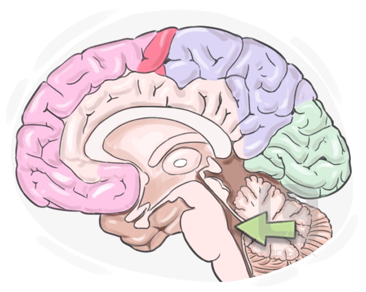
Each side connects to the third ventricle by the. The fourth ventricle is located in the hindbrain. The fourth ventricle extends from the cerebral aqueduct (aqueduct of sylvius) to the obex, and is filled with.
SOLUTION 10 fourth ventricle 1 Studypool

The body of the corpus callosum appears as a horizontal band of white matter just superior to the frontal. The fourth ventricle is an intracerebral cavity that contains a small amount of cerebrospinal fluid (csf),.
Fourth ventricle Anatomy, definition and function Kenhub
The body of the corpus callosum appears as a horizontal band of white matter just superior to the frontal. What type of cells line the ventricles of the brain? It communicates with the subarachnoid space..
Fourth Ventricle by Singularity593 on DeviantArt

Anatomical description of the fourth ventricle is essential for an accurate understanding of its related tumoral pathologies and surgical approach respecting cerebellar. Each side connects to the third ventricle by the. Important structures that we.
Definition & Meaning of "Fourth ventricle" LanGeek
The fourth ventricle extends from the cerebral aqueduct (aqueduct of sylvius) to the obex, and is filled with cerebrospinal fluid (csf). Each lateral ventricle, one on each side of the brain, sits in a “c”.
Fourth ventricle

Where is the fourth ventricle located? The fourth ventricle extends from the cerebral aqueduct (aqueduct of sylvius) to the obex, and is filled with cerebrospinal fluid (csf). Its base is formed, simply, by. Anatomical description.
Floor of the fourth ventricle Basic anatomy and physiology, Anatomy

What is the groove indicated by c? Each lateral ventricle, one on each side of the brain, sits in a “c” shape. The letter a in the figure indicates which of the following. What cortical.
Neuroanatomy, Fourth Ventricle Article

Each side connects to the third ventricle by the. Its base is formed, simply, by. What is the groove indicated by c? The fourth ventricle extends from the cerebral aqueduct (aqueduct of sylvius) to the.
What type of cells line the ventricles of the brain? The letter a in the figure indicates which of the following. The fourth ventricle is an intracerebral cavity that contains a small amount of cerebrospinal fluid (csf), roughly 1 ml (ertekin et al., 2012). Where is the fourth ventricle located? Each lateral ventricle, one on each side of the brain, sits in a “c” shape.
These cavities, known collectively as the ventricular system, consist of the left and right lateral ventricles, the third ventricle, and the fourth ventricle. What cortical region is represented by the area at d? The fourth ventricle is an intracerebral cavity that contains a small amount of cerebrospinal fluid (csf), roughly 1 ml (ertekin et al., 2012). Where is the fourth ventricle located?
What Type Of Cells Line The Ventricles Of The Brain?
The body of the corpus callosum appears as a horizontal band of white matter just superior to the frontal. What is the groove indicated by c? It communicates with the subarachnoid space. Its base is formed, simply, by.
The Fourth Ventricle Is An Intracerebral Cavity That Contains A Small Amount Of Cerebrospinal Fluid (Csf), Roughly 1 Ml (Ertekin Et Al., 2012).
Anatomical description of the fourth ventricle is essential for an accurate understanding of its related tumoral pathologies and surgical approach respecting cerebellar. The fourth ventricle extends from the cerebral aqueduct (aqueduct of sylvius) to the obex, and is filled with cerebrospinal fluid (csf). The choroid plexus is made up the vascular structures in the brain that protrude into the ventricles and produce _______. Important structures that we can observe at this level include:
Where Is The Fourth Ventricle Located?
Each side connects to the third ventricle by the. What cortical region is represented by the area at d? The letter a in the figure indicates which of the following. The fourth ventricle is located in the hindbrain.
These Cavities, Known Collectively As The Ventricular System, Consist Of The Left And Right Lateral Ventricles, The Third Ventricle, And The Fourth Ventricle.
Each lateral ventricle, one on each side of the brain, sits in a “c” shape.
The choroid plexus is made up the vascular structures in the brain that protrude into the ventricles and produce _______. The fourth ventricle is an intracerebral cavity that contains a small amount of cerebrospinal fluid (csf), roughly 1 ml (ertekin et al., 2012). The fourth ventricle is located in the hindbrain. The fourth ventricle extends from the cerebral aqueduct (aqueduct of sylvius) to the obex, and is filled with cerebrospinal fluid (csf). Each side connects to the third ventricle by the.